Full Color Illustrations

These full-color medical illustrations by freelance medical illustrator and anatomy professor Susan Decker bring anatomy, pathology, and surgical concepts vividly to life. Using dynamic color and precise detail, each piece is designed to communicate complex medical information with both scientific accuracy and visual impact. These works are created to support:

  • Medical education – illustrating anatomy, physiology, and clinical concepts for effective teaching and learning
  • Clinical reference – providing healthcare professionals with clear, detailed visuals for diagnosis, treatment, and surgical planning
  • Patient communication – helping patients understand medical conditions, procedures, and anatomy through engaging and accessible imagery

Featured subjects include detailed anatomical studies, surgical techniques, organ systems, and pathological processes—all rendered in full color to enhance depth, realism, and comprehension. Click on each image to explore more.


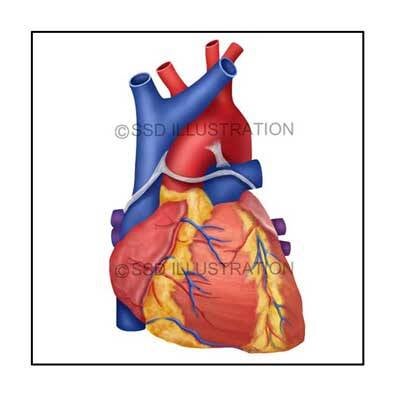
Cardiac, Gastrointestinal & Surgical Illustrations 

➤ Superficial Heart Anatomy

Full-color anatomical illustration showing the external features of the human heart, including major vessels and surface landmarks. Designed to support cardiovascular education by highlighting key structures involved in circulation and cardiac function.

➤ Gallstones

Medical illustration showing gallstones lodged in the cystic duct. Highlights anatomical relationships and clinical implications, supporting patient education, gastroenterology teaching, and surgical reference.

➤ Cardiac Valve Repair

Surgical illustration depicting heart anatomy and valve reconstruction. Designed for cardiothoracic education, surgical planning, and explaining cardiac valve repair procedures to patients and students.


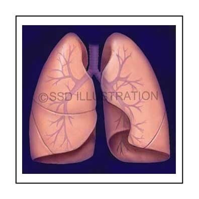
Comparative, Respiratory & Neuroanatomy Illustrations

➤ Tiger Beetle Anatomy

Detailed scientific illustration of a tiger beetle highlighting anatomical features and adaptations for predation and locomotion. Designed for entomology education, comparative anatomy studies, and to illustrate functional morphology.

➤ Human Lung Anatomy

Anatomical illustration of the human lungs showing airways and lobes. Supports medical education, respiratory system teaching, and clinical reference for lung structure and function.

➤ Dermatomes

Medical illustration mapping the distribution of spinal nerves across dermatomes. Useful for teaching neuroanatomy, explaining sensory innervation patterns, and supporting clinical discussions of nerve injury or pain localization.


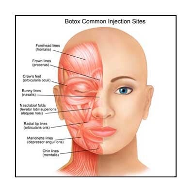
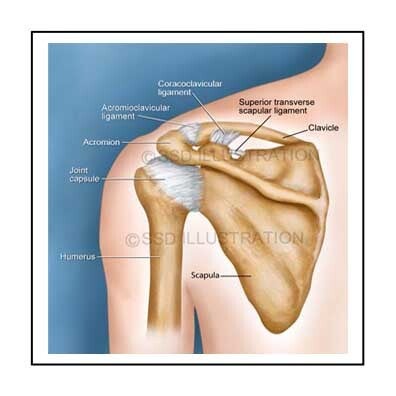
Facial, Musculoskeletal & Upper Airway Illustrations

➤ Facial Muscles and Botox

Medical illustration showing the muscles of facial expression with common Botox injection points. Created for patient education and aesthetic medicine training, highlighting anatomy relevant to facial rejuvenation and safe injection technique.

➤ Shoulder Anatomy

Anatomical illustration of the shoulder complex, depicting bones, muscles, and ligaments. Designed to clarify structural relationships for orthopedic education, physical therapy reference, and sports medicine applications.

➤ Oropharyngeal Anatomy

Anatomical illustration showing the sagittal anatomy of the mouth, throat, and pharyngeal structures. Useful for teaching upper airway anatomy, swallowing mechanics, and clinical procedures involving the oropharynx.


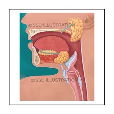
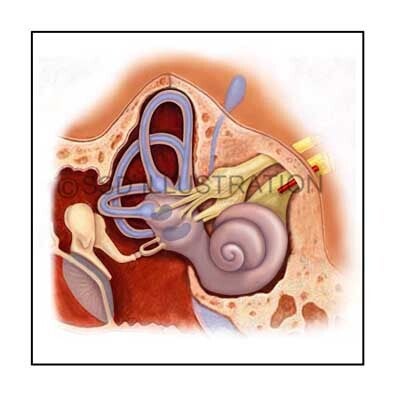
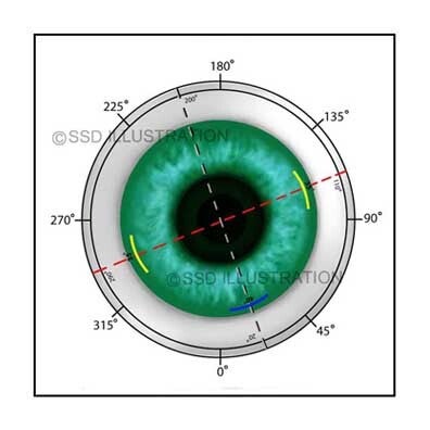
Sensory and Digestive System Illustrations

➤ Inner Ear Anatomy

Detailed anatomical illustration of the inner ear showing the cochlea, semicircular canals, and vestibular structures. Created to support otologic education by clarifying the anatomy involved in hearing and balance.

➤ Accessory Digestive Organs

Medical illustration depicting the accessory organs of the digestive system, including the liver, pancreas, and gallbladder. Designed for teaching gastrointestinal anatomy and supporting clinical understanding of digestive function and pathology.

➤ LASIK Eye Surgery

Medical illustration showing the LASIK procedure, including corneal flap creation and laser reshaping. Provides a clear visual overview for ophthalmology education, surgical reference, and patient communication about vision correction.


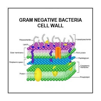
Cellular, Glandular & Molecular Illustrations

➤ Gram-Negative Bacteria

Scientific diagram showing the structure of a gram-negative bacterial cell wall, including the outer membrane, peptidoglycan layer, and lipopolysaccharide components. Designed for microbiology education to clarify bacterial architecture and antibiotic interactions.

➤ Salivary Glands and Ducts

Medical illustration of the major salivary glands labeled in relation to facial anatomy. Useful for teaching head and neck anatomy, explaining salivary function, and supporting clinical discussions of glandular disorders and surgical procedures.

➤ SREBP Cleavage

Scientific illustration depicting SREBP (sterol regulatory element-binding protein) cleavage and activation, demonstrating its role in cholesterol synthesis regulation. Created for biochemistry and molecular biology education to visualize intracellular lipid control mechanisms.