

Surgical Illustrations
These surgical illustrations by freelance medical illustrator and anatomy professor Susan Decker depict detailed operative techniques and surgical anatomy across a range of specialties. Each image is designed to support surgical education, clinical communication, and publication. Designed to support:
-
Surgical training – clarifying procedural steps and anatomical relationships for students and professionals
-
Clinical reference – providing accurate visuals for surgical planning, documentation, and consultation
-
Patient education – illustrating procedures in a clear and approachable way to improve understanding and communication
Examples include illustrations of open and minimally invasive techniques, procedural sequences, and anatomical dissections—each created to convey precision, clarity, and clinical relevance.
Surgical Illustrations: Transplant, Vascular, and Orthopedic Procedures

Pancreas Transplant Surgery
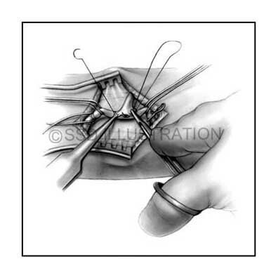
Av Fistula Surgery
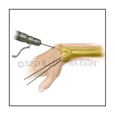
Distal Radius Repair
Scalp, Neurological & Obstetric Procedures
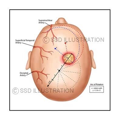
Scalp Reconstruction

Internal Uterine Massage
Trigeminal Nerve Repair
Neurological, Obstetric & Cardiac Procedures

Cardiac Valve Repair

Splenic Tumor Removal

Endoscopy and Peptic Ulcer
Robotic Surgical Access, Reconstruction & OR Configuration
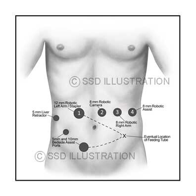
Abdominal Port Placement
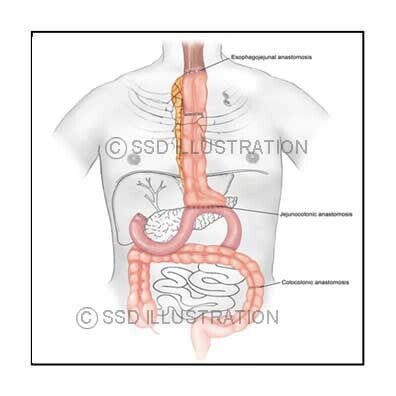
Isoperistaltic Colonic Interposition
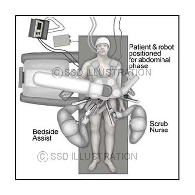
Robotic Surgery Set-up
Abdominal, Gastrointestinal & Transplant Surgery
Transplant Vessel Suturing
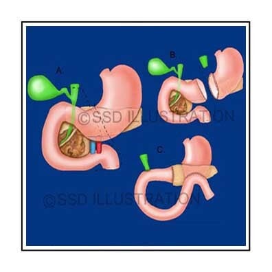
Gastric Bypass Surgery

U-Clip Placement
Ophthalmic, Orthopedic & Cardiac Surgery
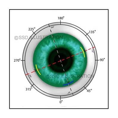
LASIK Eye Surgery

Proximal Radius Repair

Posterior Cardiac Valve Repair
