Scientific Illustrations

These scientific illustrations by freelance medical illustrator and anatomy professor Susan Decker visualize complex biological and medical concepts with clarity and precision. Each piece translates scientific data and research into clear, engaging visuals that support understanding, communication, and education.

Designed to support:

  • Research and publication – illustrating experimental methods, molecular mechanisms, and biological systems for journals and presentations
  • Education and outreach – simplifying complex science for teaching materials, lectures, and public engagement
  • Communication and visualization – transforming abstract concepts into accurate, accessible images for scientific and clinical contexts

Examples include cellular and molecular diagrams, physiological processes, and experimental workflows—each created to convey accuracy, insight, and visual impact.


Metabolism, Anatomy & Ecology Scientific Illustrations

SREBP Cleavage

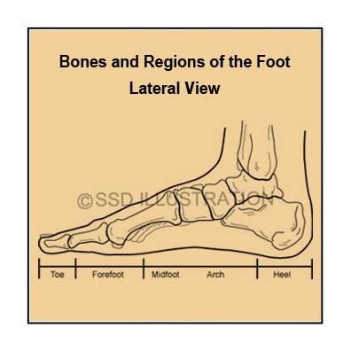
              Bones & Regions of the Foot

Hummingbird Pollination

- About These Illustrations -

  • SREBP Cleavage and Cholesterol Regulation
    Scientific illustration of SREBP cleavage showing how proteolytic activation controls cholesterol synthesis and lipid homeostasis. Ideal for use in biochemistry education, molecular biology research, and journal publication visuals.
  • Bones and Regions of the Foot
    Anatomical illustration detailing the bones and regions of the human foot, including tarsals, metatarsals, and phalanges. Designed to support anatomy education, orthopedic study, and clinical reference in podiatry and rehabilitation.
  • Hummingbird as Pollinator
    Scientific illustration of a hummingbird pollinating a flower, emphasizing coevolution and adaptations for nectar feeding. Useful for biology education, ecology presentations, and illustrating plant–pollinator relationships in environmental science.

Entomology, Muscle Physiology & Neuroanatomy Illustrations

Tiger Beetle

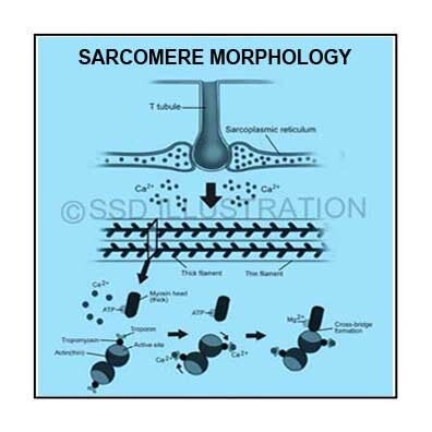
         Sarcomere Morphology

Pompano

- About These Illustrations -

  • Tiger Beetle Anatomy and Adaptations
    Detailed scientific illustration of a tiger beetle showing anatomical structures and adaptations for predation and rapid locomotion. Designed for entomology education, comparative anatomy studies, and scientific publication on insect morphology and behavior.
  • Sarcomere Structure and Muscle Contraction
    Scientific diagram illustrating sarcomere morphology, highlighting actin–myosin interactions and the mechanisms of muscle contraction. Useful for physiology courses, medical education, and visual explanations of skeletal muscle function
  • Florida Pompano 
    This scientific illustration depicts a Florida Pompano (Trachinotus carolinus) with emphasis on anatomical accuracy and species-specific coloration. Created to highlight the defining features of the species—such as its streamlined body, silvery scales, and yellow fins—this piece combines artistic rendering with scientific precision for use in educational and research contexts.


Oncology, Human Musculature & Microbiology Illustrations

Prostate Cancer Treatment

         Human Musculature

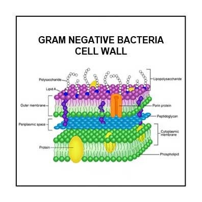
Gram Negative Bacterium

- About These Illustrations -

  • Prostate Cancer Treatment and Apoptosis
    Scientific medical illustration showing prostate cancer treatment inducing tumor cell apoptosis and preventing metastasis to the liver, spleen, and other organs. Useful for oncology education, medical publications, and patient education on treatment mechanisms and systemic effects.
  • Human Musculature Overview
    Detailed anatomical illustration of human musculature highlighting major muscle groups and their anatomical relationships. Designed for anatomy education, physical therapy reference, and medical illustration portfolios.
  • Gram-Negative Bacteria Cell Wall
    Scientific diagram depicting the structure of a gram-negative bacterial cell wall, including outer membrane, peptidoglycan layer, and lipopolysaccharide components. Ideal for microbiology education, infectious disease study, and scientific publication visuals.

Environmental Science, Pollination & Botanical Illustrations

Biomagnification

         Butterfly Anatomy

Datura Wrightii

- About These Illustrations -

  • Biomagnification from Factory Emissions
    Educational scientific illustration showing biomagnification of industrial pollutants through aquatic food chains originating from factory emissions. Designed for environmental science education, ecology studies, and illustrating human impacts on ecosystems.
  • Butterfly Head and Proboscis Anatomy
    Scientific illustration of a butterfly’s head and thorax highlighting detailed proboscis structure and adaptations for nectar feeding. Useful for pollination ecology education, entomology studies, and teaching plant–pollinator interactions.
  • Datura wrightii Morphology
    Botanical illustration of Datura wrightii showing flower morphology, leaves, and reproductive structures. Supports botany education, plant morphology studies, and scientific publication on native flora.

Parasitology, Wound Care & Infectious Disease Illustrations

Hookworm Symptoms

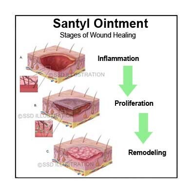
         Bed Sore Healing Progression

Tapeworm Anatomy

- About These Illustrations -

  • Hookworm Infection Symptoms
    Medical illustration showing the parasitic life cycle of hookworms, intestinal infection, and clinical manifestations. Designed for parasitology education, infectious disease reference, and patient education on helminth infections.
  • Wound Healing with Santyl Ointment
    Medical illustration of the wound healing process using Santyl collagenase ointment, showing enzymatic debridement and tissue regeneration. Useful for wound care education, nursing training, and clinical reference on advanced debridement techniques.
  • Tapeworm Anatomy
    Detailed biological illustration of tapeworm anatomy highlighting the scolex, proglottids, and internal reproductive structures. Supports parasitology education, infectious disease studies, and scientific publication visuals.