Orthopedic Medical Illustrations

These orthopedic medical illustrations by freelance medical illustrator Susan Decker depict musculoskeletal anatomy, surgical procedures, and therapeutic exercises with precision. Designed to support:

  • Medical education – clarifying complex anatomy for students and professionals

  • Clinical reference – providing detailed visuals for surgical planning and consultations

  • Patient education – simplifying complex concepts for easier understanding

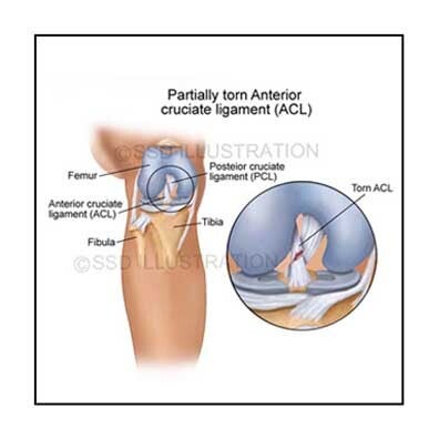
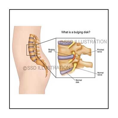
Examples include a torn ACL, shoulder physical therapy exercises, a herniated lumbar disc, and other detailed anatomical and procedural illustrations. Each image communicates complex concepts clearly for both medical professionals and patients. Click on each image to expand the selection.


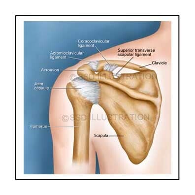
Orthopedic Anatomy – Knee, Spine & Shoulder

Torn ACL - Knee Joint

Illustration of a torn anterior cruciate ligament (ACL) in the knee, showing ligament disruption. Ideal for orthopedic consultations, patient education, and surgical planning. Helps explain knee injuries, ligament tears, and treatment options.

Shoulder Therapy Exercises

Illustration demonstrating proper technique and muscle engagement for shoulder and back exercises. Supports physical therapy, rehabilitation, and patient education. Useful for teaching correct exercise mechanics and injury prevention.

Bulging Lumbar Disk

Cross-sectional illustration of a lumbar herniated disc compressing a spinal nerve. Helps explain symptoms like sciatica and lower back pain. Useful for patient education and clinical consultations about spinal injuries and treatments.


Orthopedic Anatomy – Shoulder, Elbow & Spine

Shoulder Joint Anatomy

Detailed illustration of the shoulder’s bones and ligaments. Ideal for anatomy study, clinical reference, and patient education. Highlights structural relationships for better understanding of shoulder function and injuries..

Elbow Fracture Repair

Illustration showing fractures of the radius and common repair techniques. Supports surgical planning, orthopedic education, and patient explanation. Helps visualize treatment strategies and bone healing processes.

Lumbar Spine Anatomy

Illustration of vertebrae and intervertebral discs, highlighting spine anatomy and structure. Useful for medical education, clinical reference, and patient teaching. Clarifies spinal relationships and supports understanding of back health and injuries.


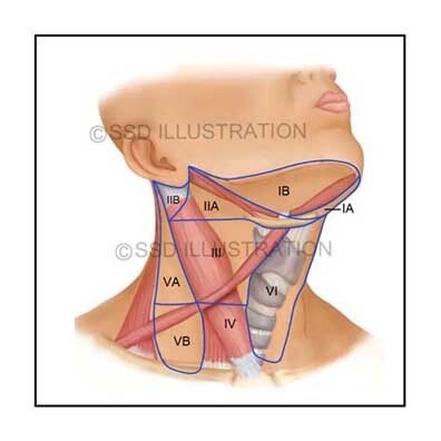
Musculoskeletal Anatomy: Radius, Pelvis & Neck
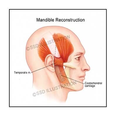
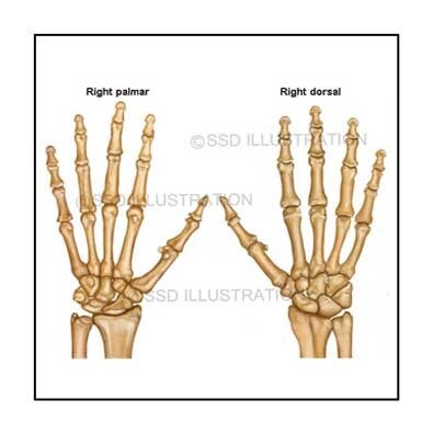
Radial Fracture Repair

Illustration showing surgical fixation techniques for a fractured radius. Supports orthopedic education, surgical planning, and patient explanation. Helps visualize bone repair and treatment strategies for upper limb injuries.

Deep Gluteal Musculature

Detailed illustration of the musculoskeletal structures of the gluteal and pelvic region. Ideal for clinical reference, anatomy study, and patient education. Highlights muscle, bone, and ligament relationships for better understanding of movement and function.

Neck Regions

Illustration of neck muscles, vertebrae, and soft tissues. Supports medical education, surgical reference, and patient teaching. Clarifies cervical anatomy for understanding injuries, conditions, and treatment options.


Orthopedic Anatomy: Foot, Hand & Knee Joint Anatomy

Foot Anatomy & Biometrics

Illustration of foot bones and their positions during running and exercise. Highlights anatomical function and biomechanics, making it ideal for medical education, physical therapy, and patient instruction.

Hand Bones & Joints

High-resolution illustration of hand bones and joints. Supports anatomy study, clinical reference, and patient education. Helps explain hand structure, function, and common injuries.

Knee Joint Anatomy

Detailed illustration of the knee joint, including bones, ligaments, cartilage, and surrounding structures. Useful for orthopedic education, clinical reference, and patient teaching. Clarifies knee anatomy for understanding injuries and treatment options.


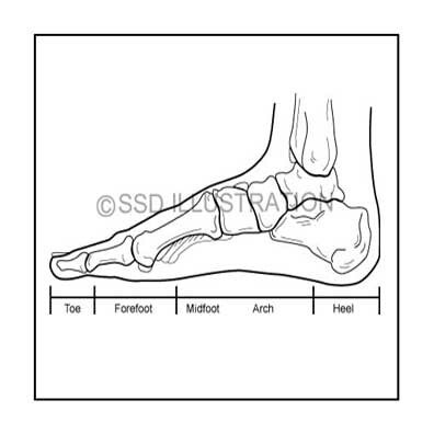
Human Musculature, Skeletal Muscle Structure, and Foot Anatomy

Human Musculature

Comprehensive illustration of major muscle groups from both front and back. Ideal for anatomy study, clinical reference, and patient education. Highlights muscular structure and relationships for a full-body understanding.

Skeletal Muscle Anatomy

Detailed illustration of skeletal muscle, showing epimysium, deep fascia, fascicles, and connective tissue. Supports scientific education, anatomy study, and clinical reference. Helps explain muscle structure and function in detail.

Foot Bones & Regions

Orthopedic illustration of the tarsals, metatarsals, and phalanges. Clarifies foot anatomy for medical education, clinical reference, and patient teaching. Highlights bone regions and structural relationships for better understanding of foot function.