

Dermatology Illustrations
These dermatology illustrations by freelance medical illustrator and anatomy professor Susan Decker explore the structure, function, and pathology of the skin. From microscopic anatomy to clinical presentations, each piece is crafted to visualize dermatologic concepts with clarity and accuracy. These works are designed to enhance:
- Medical education – illustrating skin anatomy, histology, and disease mechanisms for teaching and learning
- Clinical reference – supporting dermatologists and clinicians with precise visuals for diagnosis and treatment planning
- Patient communication – helping patients better understand skin conditions, procedures, and healing processes
Featured subjects include the layers of the skin, inflammatory and infectious diseases, wound healing, and surgical dermatology. Click on each image to view more in detail.
Facial Anatomy, Skin Cancer & Wound Care Medical Illustrations

Botox Injection Sites

Pressure Ulcer

Types of Skin Cancer
Breast Health, Esophageal Conditions & Skin Cancer Illustrations

Breast Exam Diagram

Eosinophilic Esophagitis

Melanoma on Ear & Toe
Neurology, Skin Cancer & Diabetic Neuropathy Illustrations

Dermatomes
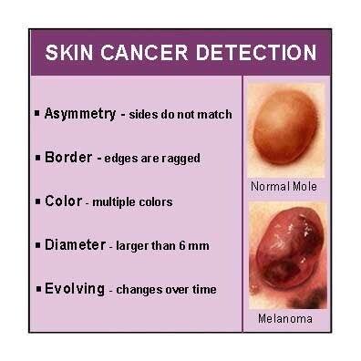
Skin Cancer Detection

Foot Screen Test Sites
Wound Care, Ear Anatomy & Skin Structure Illustrations
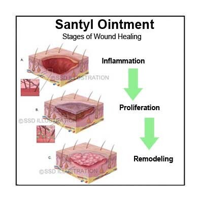
Bed Sore Healing Steps

Normal Ear Drum

Skin Layers Diagram
