Gastroenterology Illustrations

These medical illustrations of the human digestive system by freelance medical illustrator and anatomy professor Susan Decker depict digestive anatomy, physiology, pathology, and surgical procedures with precision and clarity. Each image supports medical learning, clinical communication, and publication. Designed to support:

  • Medical education – illustrating digestive anatomy, physiology, and surgical techniques for students and professionals
  • Clinical communication – providing accurate visuals for diagnosis, procedural planning, and consultation
  • Patient education – helping patients understand gastrointestinal anatomy, procedures, and conditions

Examples include anatomical diagrams, procedural sequences, and depictions of common gastrointestinal disorders—each created for accuracy, clarity, and clinical relevance. Click on each image to expand the selection.


Gastrointestinal Surgery & Diagnostic Procedures

Endoscopy For Peptic Ulcer

           Splenic Tumor Removal

Gallstone

- About These Illustrations -

  • Endoscopy Procedure: Medical illustration showing an upper gastrointestinal endoscopy with a view of the stomach lining and a visible peptic ulcer. The image highlights scope positioning, internal anatomy, and ulcer pathology for diagnostic reference.

  • Splenic Tumor Removal: Surgical illustration depicting removal of a splenic tumor with detailed anatomy of the spleen, vasculature, and surrounding organs. It emphasizes key surgical landmarks and technique relevant to splenectomy.

  • Gallstones in the Cystic Duct: Medical illustration showing gallstones obstructing the cystic duct and associated biliary anatomy. The piece illustrates the site of obstruction and its relationship to the gallbladder and hepatic ducts.


Gastroenterology & Abdominal Procedures

Pancreas Transplant Surgery

         Abdominal Regions

Eosinophilic Esophagitis

- About These Illustrations -

  • Pancreas Transplant Procedure: Surgical illustration showing the anatomical positioning of the donor pancreas and vascular connections during transplantation. Highlights key surgical landmarks and operative technique for educational reference.

  • Abdominal Regions: Medical illustration depicting the nine labeled abdominal regions, designed for patient education. Shows surface anatomy landmarks used in gastroenterology to identify organ locations and abdominal pain areas.

  • Stages of Eosinophilic Esophagitis: Medical illustration showing progressive esophageal inflammation and tissue changes. Demonstrates disease progression and anatomical impact for both clinical and patient education purposes.


Digestive System, Splenic Injuries & Gastric Bypass

Splenic Injury Grading

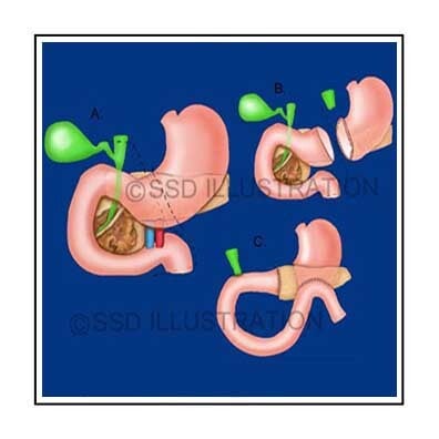
         Gastric Bypass Surgery

Digestive System Anatomy

- About These Illustrations -

  • Splenic Injury Grading: Medical illustration showing different levels of spleen trauma, highlighting anatomical structures and injury severity for clinical reference and surgical education.

  • Gastric Bypass Procedure: Surgical illustration detailing the gastric bypass procedure, including anatomical changes, operative steps, and key surgical landmarks for educational purposes.

  • Digestive System Anatomy: Anatomical illustration of the human digestive system showing major organs, including the stomach, liver, pancreas, spleen, and intestines, designed for medical and patient education


Digestive System & Accessory Organ Anatomy

Salivary Glands & Ducts

  Liver Segments

Digestive Accessory Organs

- About These Illustrations -

  • Salivary Glands: Medical illustration showing the major salivary glands labeled in relation to facial anatomy, highlighting their positions and relationships for educational and clinical reference.

  • Liver Segments: Medical illustration depicting liver segments with surrounding digestive system anatomy labeled, emphasizing anatomical organization and functional regions for teaching and clinical planning.

  • Accessory Digestive Organs: Medical illustration of the accessory organs of the digestive system, including liver, pancreas, and gallbladder, showing their anatomical positions and relationships for medical and patient education.