Medical Illustrations

These medical illustrations by freelance medical illustrator and anatomy professor Susan Decker depict anatomical structures, clinical procedures, and pathological conditions. Each image is designed to support medical education, clinical reference, and patient communication. Designed to support:

  • Medical education – clarifying complex anatomy for students and professionals

  • Clinical reference – providing detailed visuals for surgical planning and consultations

  • Patient education – simplifying complex concepts for easier understanding

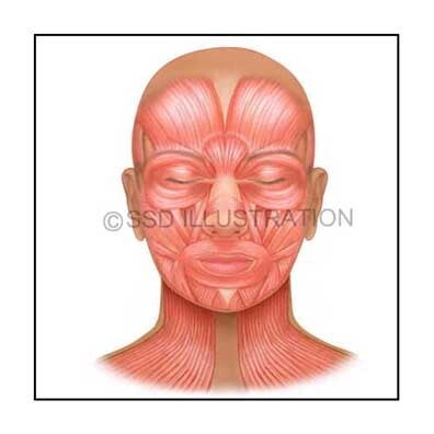
Examples include illustrations of facial muscles, the circulatory system, and various organ systems, each crafted to enhance comprehension and communication in medical settings. Click on each image to expand the selection.


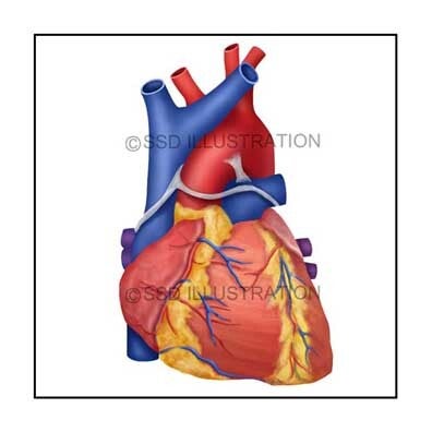
Cardiovascular, Neurological, & Spinal Anatomy

External Heart Anatomy

Full-color anatomical illustration showing the external features of the human heart, including major vessels and surface landmarks. Designed to support cardiovascular education by highlighting key structures involved in circulation and cardiac function.

Petrous Bone Repair

A detailed surgical illustration showing the trigeminal nerve in relation to surrounding cranial structures, with emphasis on the exposure and repair of the petrous portion of the temporal bone to support clinical and educational visualization.

Lumbar Spine Anatomy

An anatomical illustration depicting the lumbar vertebrae and sacrum, highlighting key structural features of the lower spine for musculoskeletal education and clinical reference.


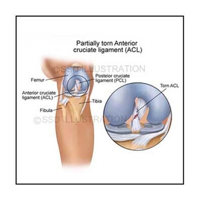
Clinical Anatomy: Knee, Gallbladder & Throat

Torn ACL - Knee Joint

An orthopedic medical illustration showing a ruptured anterior cruciate ligament, with clear visualization of surrounding joint structures to support surgical planning and patient education.

Gallstones

A medical illustration demonstrating gallstones obstructing the cystic duct, emphasizing biliary anatomy and common sites of blockage for diagnostic and educational use.

Regions of the Neck

A diagram illustrating the anatomical quadrants of the neck with labeled regions, designed to clarify spatial organization for clinical examination and instructional purposes.


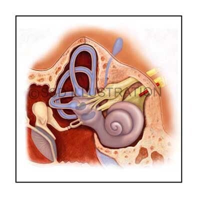
Urogenital and Ear Anatomy Illustrations

Uterine Remnant in Male

A sagittal anatomical illustration of the male reproductive and urinary organs, including the visualization of a uterine remnant in a male child, created for medical education and clinical reference.

Inner Ear Anatomy

A detailed depiction of the cochlea and semicircular canals, highlighting key structures involved in hearing and balance for anatomical study and clinical instruction.

Male Urogenital System

An anterior anatomical illustration of the male reproductive and urinary organs, designed to support educational use, clinical teaching, and anatomical reference.


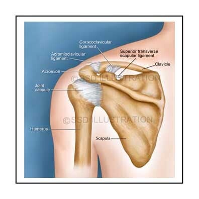
Musculoskeletal, Dermatological, & Facial Anatomy

Shoulder Joint Anatomy

An anatomical illustration of the shoulder joint highlighting major bones, muscle groups, and ligament structures, designed to support clinical understanding and medical education.

Melanoma – Ears and Toes

A medical illustration depicting melanoma lesions on the ear and toes, emphasizing visual presentation and location to aid in clinical teaching and patient awareness.

Facial muscles – Anatomical Study

An illustration detailing the major muscle groups of the face, providing clear structural organization for anatomical learning and reference.


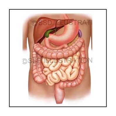
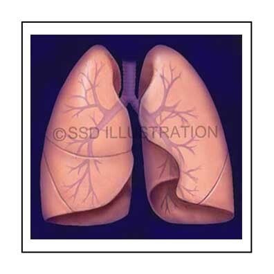
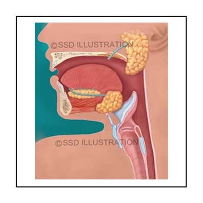
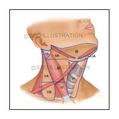
Digestive, Respiratory, & Pharyngeal Anatomy

Digestive System Anatomy

A comprehensive anatomical illustration of the major digestive organs, including the stomach, intestines, liver, and accessory structures, designed to support detailed anatomical education and clinical reference.

Lung - Lobes & Airways

A detailed depiction of the human lungs, highlighting the bronchial tree, lobes, and major airways, created to enhance understanding of respiratory anatomy for medical study and clinical applications.

Oropharynx - Sagittal View

A sagittal anatomical illustration showing the oral cavity, pharynx, and related structures, emphasizing spatial relationships and anatomical landmarks for educational and clinical study.